Cervical Spine X Ray / Cervical Spine X-ray | Cervical Spine -- Left Anterior ... - You want to see the entire cervical spine so that you can make sure that there is not an injury there.
Cervical Spine X Ray / Cervical Spine X-ray | Cervical Spine -- Left Anterior ... - You want to see the entire cervical spine so that you can make sure that there is not an injury there.. Cervical spine radiographic series contains 3 views. Awareness of the prevalence of incidental findings is important in. Normal cervical spine radiographs in a young adult. Several injury patterns can widen this space. They may be taken to find injuries or diseases that affect the discs or joints in your spine.
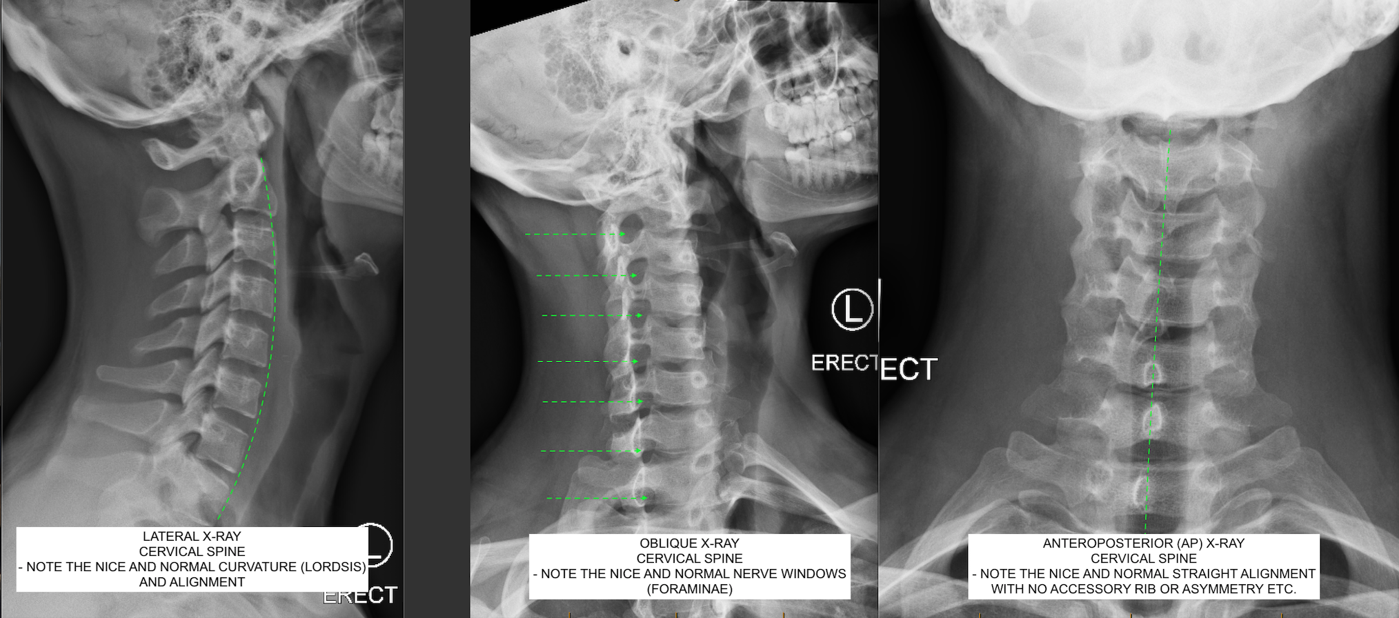
Fracture of the ring of c1 can allow the anterior portion of the ring to migrate anteriorly with respect to the dens. You require all three views (lateral, ap and odontoid/open mouth view) for an adequate study. Cervical spine pa or ap. The density should be appropriate with soft tissues and bony structures well visualized. The standard three views taken are the
Dynamic cervical spine X-ray in a patient who underwent 2 ... from www.researchgate.net Make sure you can see all 7 cervical spinous process. You require all three views (lateral, ap and odontoid/open mouth view) for an adequate study. See canadian cervical spine rule. Incongruencies indicate mainly cervical fracture, spondylolisthesis or ligament injury. Lying down method is done in cases of injury or patients who cannot stand. Odontoid/pedicle fracture of c2 (unstable) 8. The first method is done routinely on ambulatory patients in the outpatient department. Fracture of the ring of c1 can allow the anterior portion of the ring to migrate anteriorly with respect to the dens.
You require all three views (lateral, ap and odontoid/open mouth view) for an adequate study.
Cervical spine pa or ap. Normal cervical spine radiographs in a young adult. Lying down method is done in cases of injury or patients who cannot stand. Sacral area (base of the spine). Incongruencies indicate mainly cervical fracture, spondylolisthesis or ligament injury. Other related procedures that may be used to diagnose spine, back, or neck problems include myelography (myelogram), computed tomography (ct scan), magnetic resonance. See canadian cervical spine rule. Look for alignment of four parallel vertical columns that follow a slightly lordotic curve without any step offs. The high complexity of the anatomical structure of the craniovertebral zone explains the need to identify the main radiographic guidelines used in its evaluation. The density should be appropriate with soft tissues and bony structures well visualized. Fracture of the ring of c1 can allow the anterior portion of the ring to migrate anteriorly with respect to the dens. Make sure you can see all 7 cervical spinous process. Advantages •can quickly identify if a fracture or other suspected bony pathology is.
It shows cervical vertebra one through five. Odontoid/pedicle fracture of c2 (unstable) 8. You want to see the entire cervical spine so that you can make sure that there is not an injury there. The first method is done routinely on ambulatory patients in the outpatient department. Advantages •can quickly identify if a fracture or other suspected bony pathology is.
cervical vertebrae x ray - Google Search | Cervical ... from i.pinimg.com Sacral area (base of the spine). So what are we looking at? They may be taken to find injuries or diseases that affect the discs or joints in your spine. This post is real simple. Most spinal injuries occur at the junctions of the spine. Cervical spine radiographic series contains 3 views. The density should be appropriate with soft tissues and bony structures well visualized. Learn vocabulary, terms and more with flashcards, games and other study tools.
When reading any radiograph the clinician should establish a process or order they follow each time.
A specially trained technician will position you on the table so that the section of your spine getting. The first method is done routinely on ambulatory patients in the outpatient department. Odontoid/pedicle fracture of c2 (unstable) 8. Look for alignment of four parallel vertical columns that follow a slightly lordotic curve without any step offs. Several injury patterns can widen this space. Most spinal injuries occur at the junctions of the spine. Lying down method is done in cases of injury or patients who cannot stand. The high complexity of the anatomical structure of the craniovertebral zone explains the need to identify the main radiographic guidelines used in its evaluation. See canadian cervical spine rule. Other related procedures that may be used to diagnose spine, back, or neck problems include myelography (myelogram), computed tomography (ct scan), magnetic resonance. You require all three views (lateral, ap and odontoid/open mouth view) for an adequate study. They may be taken to find injuries or diseases that affect the discs or joints in your spine. Thoracic spine (chest or trunk area).
You want to see the entire cervical spine so that you can make sure that there is not an injury there. Normal cervical spine radiographs in a young adult. So what are we looking at? The standard three views taken are the Advantages •can quickly identify if a fracture or other suspected bony pathology is.
C.N.S. Neurosurgery | Normal Cervical Spine X-ray from www.cnsneurosurgery.com.au Advantages •can quickly identify if a fracture or other suspected bony pathology is. See canadian cervical spine rule. Fracture of the ring of c1 can allow the anterior portion of the ring to migrate anteriorly with respect to the dens. So what are we looking at? The high complexity of the anatomical structure of the craniovertebral zone explains the need to identify the main radiographic guidelines used in its evaluation. This post is real simple. Several injury patterns can widen this space. When reading any radiograph the clinician should establish a process or order they follow each time.
This post is real simple.
A specially trained technician will position you on the table so that the section of your spine getting. The high complexity of the anatomical structure of the craniovertebral zone explains the need to identify the main radiographic guidelines used in its evaluation. Odontoid/pedicle fracture of c2 (unstable) 8. Case contributed by dr andrew dixon ◉. You require all three views (lateral, ap and odontoid/open mouth view) for an adequate study. Sacral area (base of the spine). See canadian cervical spine rule. You want to see the entire cervical spine so that you can make sure that there is not an injury there. The standard three views taken are the Most spinal injuries occur at the junctions of the spine. Learn vocabulary, terms and more with flashcards, games and other study tools. Lying down method is done in cases of injury or patients who cannot stand. It shows cervical vertebra one through five.
You have just read the article entitled Cervical Spine X Ray / Cervical Spine X-ray | Cervical Spine -- Left Anterior ... - You want to see the entire cervical spine so that you can make sure that there is not an injury there.. You can also bookmark this page with the URL : https://ndankbar.blogspot.com/2021/05/cervical-spine-x-ray-cervical-spine-x.html
Share Awesome
Belum ada Komentar untuk "Cervical Spine X Ray / Cervical Spine X-ray | Cervical Spine -- Left Anterior ... - You want to see the entire cervical spine so that you can make sure that there is not an injury there."


Belum ada Komentar untuk "Cervical Spine X Ray / Cervical Spine X-ray | Cervical Spine -- Left Anterior ... - You want to see the entire cervical spine so that you can make sure that there is not an injury there."
Posting Komentar